When Jeremy Piven abruptly quit Speed The Plow a while back, saying that his blood mercury level was 57 (10 is the upper limit of normal) had left him too ennervated to perform, the producers cried foul. Given various reports of hard partying, the producers felt Piven was using this as an excuse to quit the show. The wittiest line to emerge out of the whole brouhaha came from the play’s author, David Mamet.
“I talked to Jeremy on the phone, and he told me that he discovered that he had a very high level of mercury,” Mamet said. “So my understanding is that he is leaving show business to pursue a career as a thermometer.”
But a recent blurb in eatocracy reminded us that we never really understood the neuroscience of mercury poisoning – the chief symptom of which is fatigue – in the first place.
To answer this question we need to connect at least two dots. First, how does mercury affect the brain? And second, how does a brain produce fatigue?
Mercury in the Brain
In fish, mercury destorys or prevents the development of serotonergic neurons in the hypothalamus (Tsai). It also reduces the degradation of serotonin by inactivating MAO-B in humans who eat high-mercury fish and have blood mercury levels above 3.4 mcg/L (75th percentile – Piven was way above this). However other studies indicate that mercury may increase serotonin loss.
The Serotonin Story
In invertebrates – the sorts of animals Piven was apprently fond of eating – serotonin is a sort of master controller of energy balance, by regulating a host of central pattern generators (CPGs) involved in foraging for food, ingesting food, and storing the energy that thereby results.
Take this rather gripping anecdote from Tecott’s article (reference below).
“Hirudo is a segmented worm-like creature possessing an anterior sucker that contains a mouth with a set of tripartite jaws. Hungry leeches patrol the surface of ponds and swim toward the source of disturbances in the water (Lent et al., 1991). When they encounter warm surfaces, they bite using their three serrated jaws, and rhythmic pharyngeal contractions direct the blood meal into the digestive organ (crop) (Lent, 1985). A single feeding episode may increase their mass 8-fold (Lent et al., 1988), followed by a prolonged period of satiation (up to 1 year). Satiated leeches remain relatively lethargic and seek cover in deep water. Many aspects of these processes are subject to regulation by massive (50–100 mm diameter) serotonergic neurons (Retzius cells) that reside in pairs within each of the 32 ganglia comprising the animal’s segmented nervous system. The serotonin content of these neurons reflects their state of satiation (Lent et al., 1991). Typically, hungry leeches have high Retzius cell serotonin levels. These levels drop during feeding (Lent, 1985) and remain low during the period of satiation. In accord with this, pharmacological depletion of serotonin reduces both approach behavior and biting directed toward food-related stimuli (Lent and Dickinson, 1984). Conversely, leeches treated with serotonin will consume larger meals, and recently sated leeches may be induced to resume biting in the presence of serotonin (Lent, 1985). Retzius neuron firing rates increase when the lip of the anterior sucker contacts a warm surface (Groome et al., 1995). These neurons innervate the jaws, pharynx, salivary glands, and body wall musculature, where they stimulate biting, pharyngeal contractions, production of saliva (containing the anticoagulant hirudin), and body wall muscle relaxation permitting expansion of the crop. Crop distension, in turn, has been shown to suppress feeding behavior, ganglionic serotonin content, and Retzius cell firing rates (Groome et al., 1995). Thus, these serotonergic neurons respond to both external and internal ingestion-related stimuli to coordinate diverse responses to food.”
Vertebrate – such as Piven himself – are the opposite: serotonin dampens, rather than increases feeding – as those familiar with the rumor that tryptophan, the precursor to serotonin, is high in turkey, and is the cause of the ‘full’ feeling one gets after a Thanksgiving meal. A smaller number of readers may recall that Fen-Phen (fenfluramine) helped people diet by boosting brain serotonin and reducing appetite.
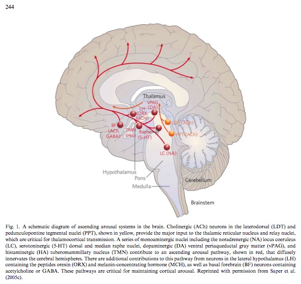
Athough the serotonin produced in the dorsal raphe nucleus (DRN) projects to virtually every forebrain structure involved in feeding, it is likely that a major mechanism of serotonin’s suppression of appetite is its effect on the hypothalamus, the ‘master controller’ of feeding behavior. Serotonin injected into the paraventricular nucleus of the hypothalamus (PVN), as well as the dorsomedial (DMN) and ventromedial (VMN) nuclei reduce feeding.
References
Saper. Staying awake for dinner: hypothalamic integration of sleep, feeding, and circadian rhythms. Prog Brain Res (2006) vol. 153 pp. 243-52
Tecott. Serotonin and the orchestration of energy balance. Cell Metab (2007) vol. 6 (5) pp. 352-61
Tsai et al. Effects of mercury on serotonin concentration in the brain of tilapia, Oreochromis mossambicus. Neuroscience Letters (1995) vol. 184 (3) pp. 208-11
Stamler et al. Relationship between platelet monoamine oxidase-B (MAO-B) activity and mercury exposure in fish consumers from the Lake St. Pierre region of Que., Canada. Neurotoxicology (2006) vol. 27 (3) pp. 429-36
Abdelouahab et al. Monoamine oxidase activity in placenta in relation to manganese, cadmium, lead, and mercury at delivery. Neurotoxicology and Teratology (2009) pp.


Hello there, You’ve performed a great job. I’ll certainly digg it and in my opinion recommend to my friends. I’m sure they will be benefited from this website.
Magnificent goods from you, man. I have understand
your stuff previous to and you’re just too excellent.
I actually like what you have acquired here, really like what you
are saying and the way in which you say it. You make it enjoyable and you still
take care of to keep it smart. I can not wait to read far more from you.
This is actually a great web site.